Over 35 years experience
YOUR DENTISTS IN
PLATEAU MONT-ROYAL
The distinguishing feature of our clinic is its pioneering use of technology. In addition to paperless management of dental records, numerous technologies allow us to provide dental care comfortably, quickly and with great precision.
Digital X-Rays 2D and 3D
Digital X-Rays
2D and 3D
In our dental clinic, digital x-rays have replaced traditional x-ray films. In addition to making it easier to store images in patient records, this new technology also comes with other benefits:
- 90% reduction in exposure to radiation (as opposed to traditional x-rays).
- Immediate results – meaning less waiting time for you.
- More eco-friendly, thanks to the elimination of toxic solutions used to process traditional x-ray films.
- Quicker transfer of images, as x-rays can be sent by email to insurance companies, dentists and other medical professionals.
- The digital images are processed by computer to improve the contrast and luminosity. This allows improved controls and often prevents taking additional x-rays when images are not clear enough.
Types de radiographies
dentaires
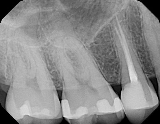
Periapical x-rays show a group of upper or lower teeth so as to better examine the root, crown and supporting bone structure of the teeth.
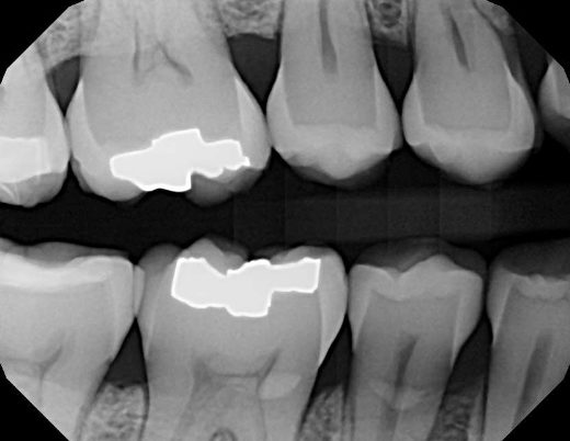
Interproximal x-rays, also known as bitewings, show the crowns of the upper and lower teeth; it helps the dentist to detect cavities between the teeth at the back of your mouth and evaluate the level of the supporting bone structure.
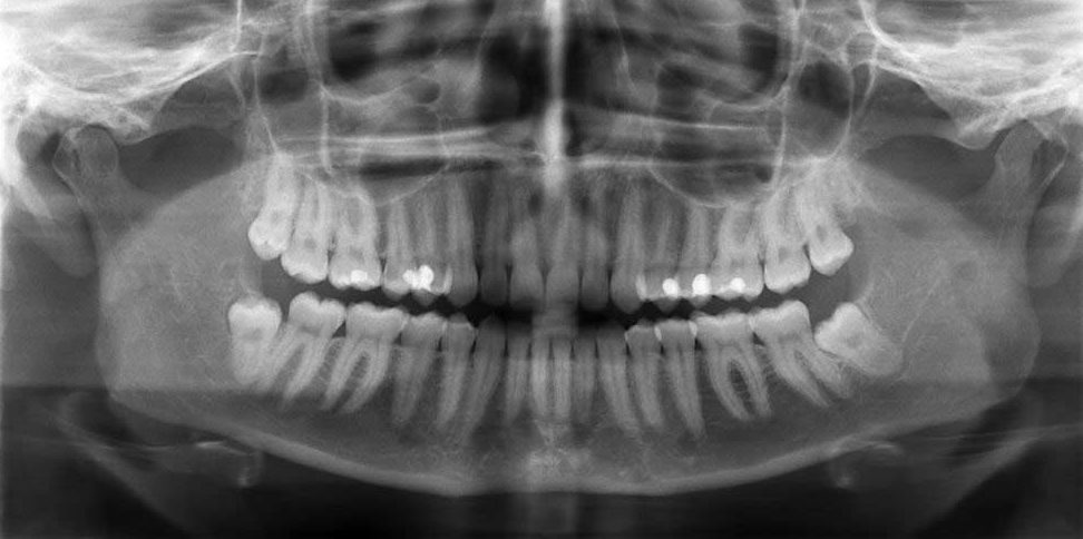
Interproximal x-rays, also known as bitewings, show the crowns of the upper and lower teeth; it helps the dentist to detect cavities between the teeth at the back of your mouth and evaluate the level of the supporting bone structure.
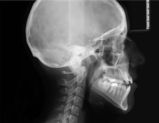
The cephalogram is an x-ray of the skull and jaws. It is mainly used in orthodontics to evaluate bone growth and teeth development.
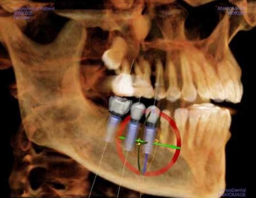
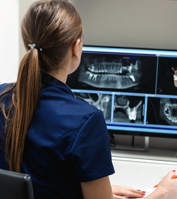
Cone Beam Computed Tomography (CBCT) is the most advanced type of dental x-rays and is available in very few clinics. The image produced is in 3 dimensions. In the above example, it is used in the precise planning of implant placement.
Contact - EN
"*" indicates required fields

