Thanks to constant research and technological development, dentists now have even more tools at their disposal. Existing technologies are becoming more precise and easier to use. Among them are two-dimensional and three-dimensional dental X-rays, essential tools for diagnosis and treatment planning. Here is an overview.
Digital X-rays: Why Use Them?
X-rays, which are internal images produced using X-ray beams, have long been used in both general and dental medicine. In dentistry, this tool allows practitioners to see structures of the mouth and skull that are otherwise invisible. This includes bones, dental roots, periodontal tissues, and teeth that are still inside the jaw (wisdom teeth or developing permanent teeth). Dentists can assess their condition, their position, and detect any signs of pathology.
Digital X-ray devices, like those used at Centre Dentaire de Haute Technologie du Québec in Montreal, are completely safe and emit 80 to 90 percent less radiation than traditional X-rays. They can be used on people of all ages, except pregnant women who are generally advised to avoid them.
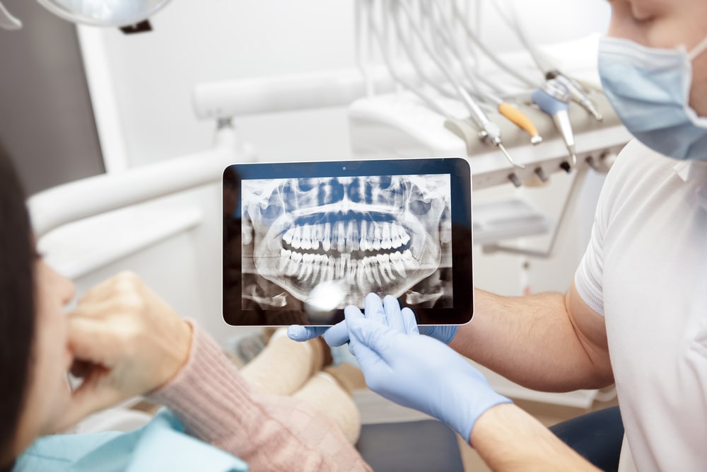
Two-Dimensional and Three-Dimensional Dental X-rays: Two Technologies, Different Results
Two-Dimensional X-rays
These versatile images allow dentists to examine a specific area of the mouth or the entire dentition to identify a variety of issues.
- Periapical and interproximal X-rays: Isolate a specific group of teeth (upper, lower, or both). Periapical X-rays show the roots, crowns, and supporting bone. Interproximal X-rays are useful for detecting cavities between the back teeth.
- Panoramic X-rays: Provide an overall view of the mouth and also show skull structures such as the maxilla, mandible, sinuses, and temporomandibular joint.
- Cephalometric X-rays: Provide a side view of the face, useful for tracking the development and position of oral and facial structures.
Three-Dimensional X-rays
Also known as cone beam volumetric tomography, these images produce a three-dimensional view of the jaw bones and surrounding tissues (roots, nerves, blood vessels, sinuses). They are used to evaluate shape, development, and specific parameters such as bone depth and density or root morphology.
Dental X-Rays: An Exceptional Diagnostic Tool
Because they allow dentists to see structures that are invisible during a regular visual exam, X-rays—particularly two-dimensional ones—serve as a valuable complement. They help dentists identify problems occurring beneath the gums or refine the diagnosis of partially visible conditions. For example, they help determine the extent of a cavity located on the root or along the side of a tooth.
A Technology for Treatment Planning
Both two-dimensional and three-dimensional X-rays are essential for evaluating the feasibility or necessity of certain procedures, planning treatment steps, and assessing outcomes.
Dental Implants
Before placing implants, both two-dimensional and three-dimensional X-rays are taken. They allow dentists to analyze bone density and thickness, determine whether a bone graft is needed, and identify the ideal location and depth for the implant screws.
Tooth Extractions
X-rays are used to evaluate the position and potential issues related to wisdom teeth, and to determine whether they should be removed preventively. X-rays also help anticipate extraction difficulty and plan surgical steps.
Orthodontic Treatments
Radiographic images are used to identify the desired changes and monitor progress throughout the treatment, particularly using cephalometric (side view) images.
Given the many functions of dental X-rays, it is very likely that your dentist in Plateau Mont Royal will use them during one of your next visits. If you have any questions, feel free to ask. We will be happy to reassure you about the safety of this technology.